Brain Surgery
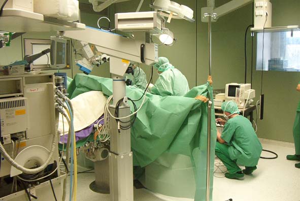
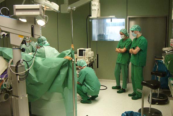
As part of my Master's project, I had the opportuniy to observe an operation to remove a brain tumor. The patient had experienced some personality changes, and was having trouble speaking.
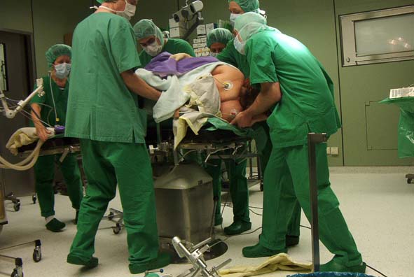
The patient was already anesthetized when we arrived.
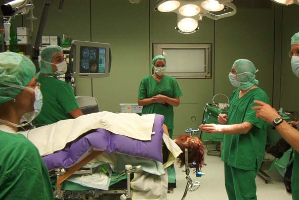
First step was to calibrate the sensing equipment. The equipment was from Surgical
Navication Technologies, a Denver firm where I had my first ever job interview.
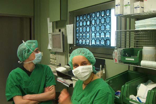
Frank and Crisi check out the 3D X-ray images while we wait for the action to begin.
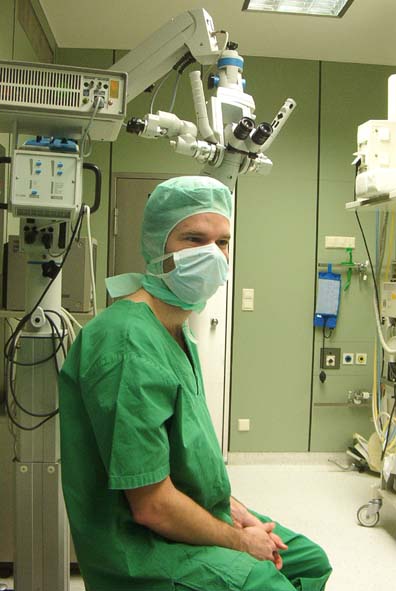
Somehow I got the extra-cool full wrap-around surgeon-style hair covering.
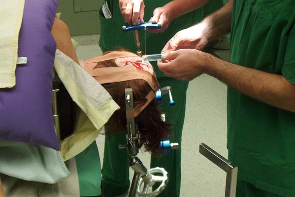
The first cut is made, and local anesthetic is injected.
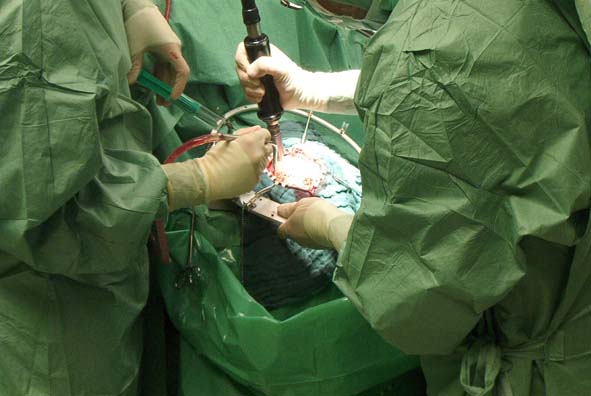
A special drill is used that will cut through bone, but will not harm soft tissue. (Like, for example, a brain.)
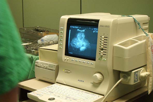
In adition to the fancy SNT system, old-fashioned ultrasound is also used.
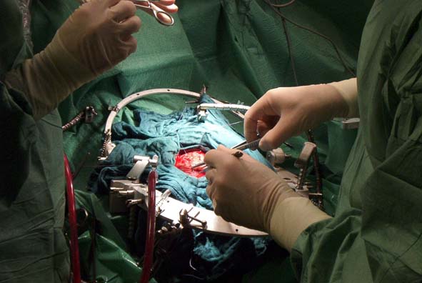
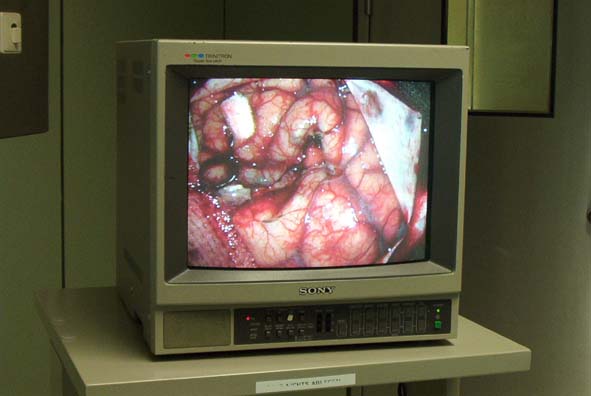
When a hospital photographer came in for a few shots, they temporarily turned off the blinding spotlights which allowed for a few good shots of the brain.
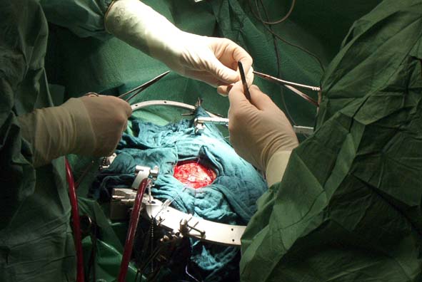
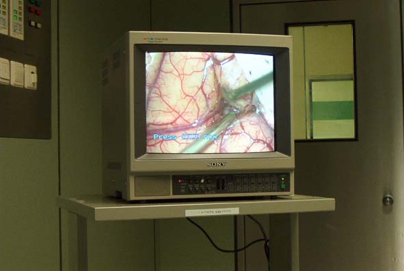
Once the surgeon got to work digging down to the tumor, all the action could be seen on this TV monitor.
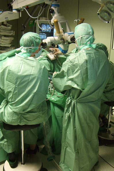
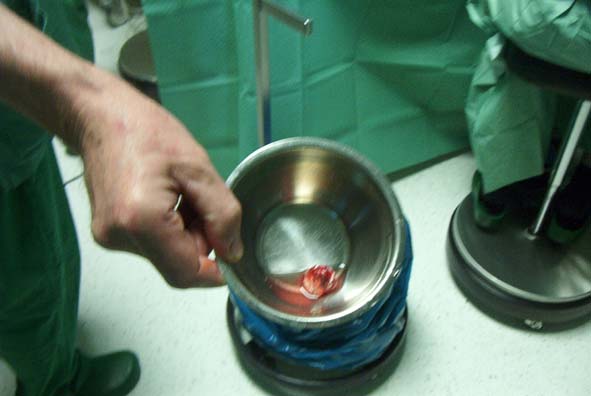
And finally, the tumor was removed.
Removing the tumor was only the halfway point. There was still over two hours of surgery to go, putting everything back together.
